A coating inspired from the cell membrane: LipoCoat® in three applications
New LipoCoat® technology brochure
Fabian Stein
September 29, 2026

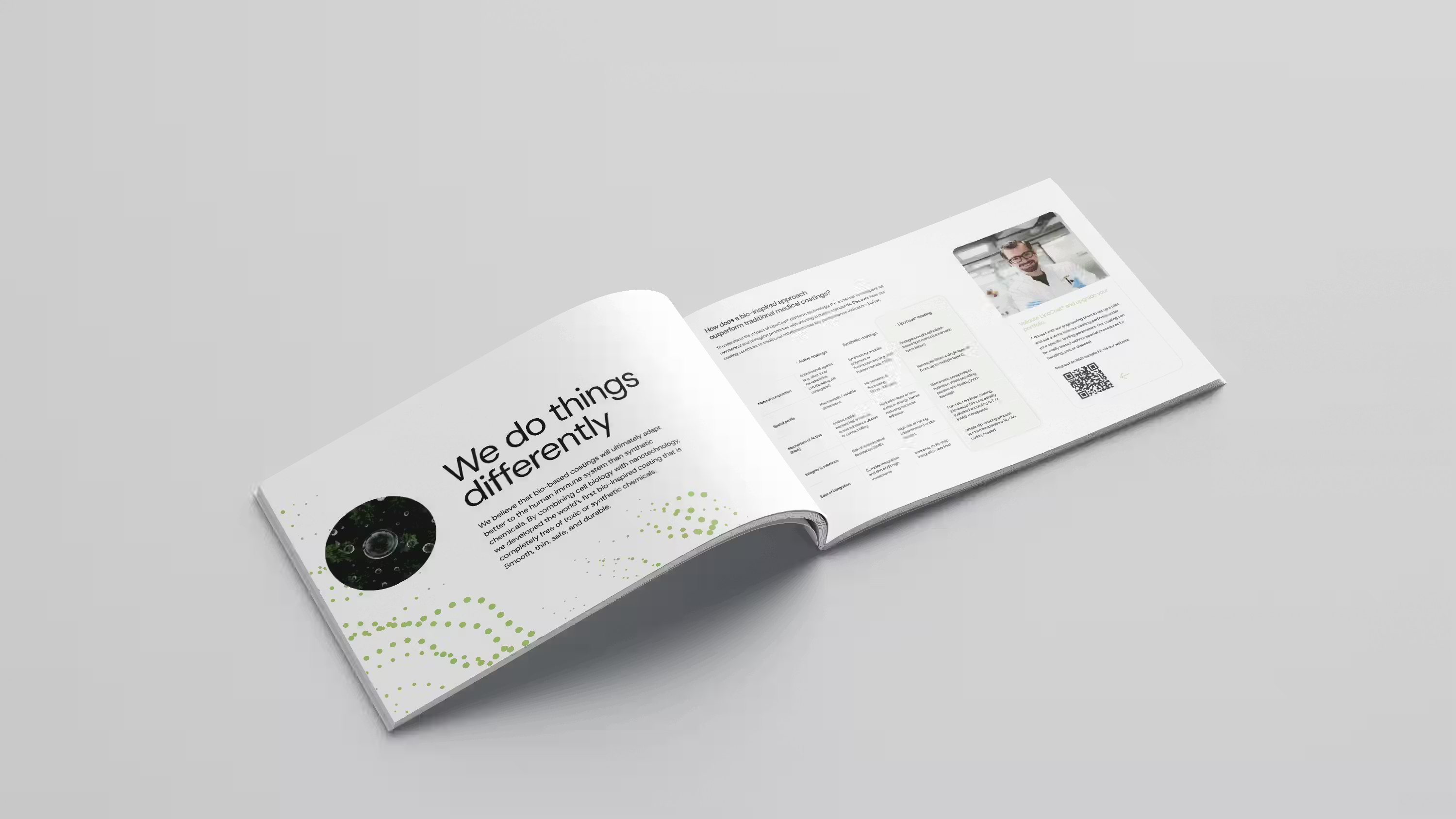
Medical device coatings have long forced a trade-off: kill bacteria and risk resistance, or add thick synthetic layers that can flake. Our new LipoCoat® technology brochure explains a third route, a nanoscale lipid layer modeled on the human cell membrane, and shows where it is already on the market.
Every device that enters the body meets the same biology. Within minutes, proteins settle on the surface, bacteria follow, and the body starts treating the device as a foreign object. The brochure describes how LipoCoat® interrupts that sequence at the very first step, and what that means for three device categories.
LipoCoat® is built from endogenous phospholipids, the same class of molecules that forms the outer membrane of human cells. Their hydrophilic heads attract water and bind it into an extremely smooth, slippery layer on the device surface. Proteins and bacteria find nothing to hold on to. Because the surface resembles the body's own tissue, the coating also mitigates the foreign body response that normally follows insertion or implantation.
The layer is nanoscale: a single layer measures 5 nm, and multiple layers can be built up where an application needs more. The formulation contains no active pharmaceutical ingredients (APIs), no silicone and no PFAS.
The brochure sets LipoCoat® against the two coating families that dominate the market today.
For cardiovascular and urological catheters, LipoCoat® is applied at around 500 nm for single-use devices that stay in place for up to 30 days. Under static conditions (modified ASTM E3151) the coating reduced bacterial adhesion by 99%. The more demanding test ran central venous catheters (CVCs) in an in vitro blood flow model at a shear stress of 7.5 dyn/cm² for up to 29 days. Uncoated controls developed dense microbial colonization. The LipoCoat®-treated catheters stayed free of biofilm for the full period.
The coating also limits protein adsorption, which plays a role in thrombus formation, and lowers friction to reduce tissue trauma during insertion. This application is on the market: the CEMMA CVC catheter with LipoCoat® received TFDA approval.
On reusable contact lenses worn for up to six months, a single 5 nm layer holds a stable, continuous film of moisture across the lens throughout the day. That wettability improves comfort and reduces mechanical friction with every blink. The coating also resists protein deposits and surface contamination, and it binds and stabilizes lysozyme, the eye's own antibacterial enzyme. It offers a non-toxic alternative to standard lubricants, and this application is on the market as well.
Injection and aspiration needles are traditionally siliconized to ease insertion. According to the brochure, LipoCoat® delivers immediate dry friction reduction comparable to silicone, and outperforms conventional silicone oil on stainless steel needles under wet conditions. Lower insertion force means more comfort for the patient, with no toxic lubricant involved. Needle applications are currently in development.
LipoCoat® is applied with the Fast Amphiphile Solution Transfer (FAST) method: clean, dip once, dry. The process runs at room temperature and needs no heating, UV curing or primer. Layer thickness is determined by the molecular properties of the coating, so the result stays uniform regardless of how the dip is performed. The step fits into existing manufacturing lines without major modifications or specialized tooling, and material consumption is minimal.
The regulatory path is simpler as well. As a passive barrier with no active substances, the coating requires no pharmaceutical classification. Biocompatibility has been evaluated under ISO 10993-1, production runs under ISO 13485:2016 in our 1,000 m² facility in Enschede, and all compliance documentation is supplied in digital format. The coating is fully biodegradable and produces non-halogenated waste streams.
The performance data in the brochure comes from in vitro testing: static adhesion assays, a dynamic blood flow model, and friction and contact angle measurements. Those tests show how the surface behaves. They do not report infection rates, thrombosis rates or patient outcomes, and they were run on specific substrates and geometries.
Your device has its own polymer, its own shape and its own dwell time in the body. The way to find out how LipoCoat® performs on it is to coat it and test it against your own specifications. That is what our R&D sample kits and pilot projects are for, backed by in-house surface, microbial, friction and biocompatibility testing aligned with ASTM and ISO standards. Talk to our engineering team to set one up.
The LipoCoat® technology brochure contains the full comparison table, the specifications per application and the test conditions behind these results. Download the brochure here.


See the specifications, test data and integration steps behind a 5 nm bio-inspired coating.
LipoCoat and SonoCoat have earned recognition from leading institutions and regulatory bodies. Our coatings meet the highest standards for biocompatibility and clinical efficacy.

LipoCoat is building a 500-square-meter production lab across from the University of Twente and is marking its tenth anniversary. Constantijn van Oranje started construction.

Four independent peer-reviewed studies have evaluated SonoCoat®, from EUS-FNA needles to prostate brachytherapy. What each one tested, and what it found.
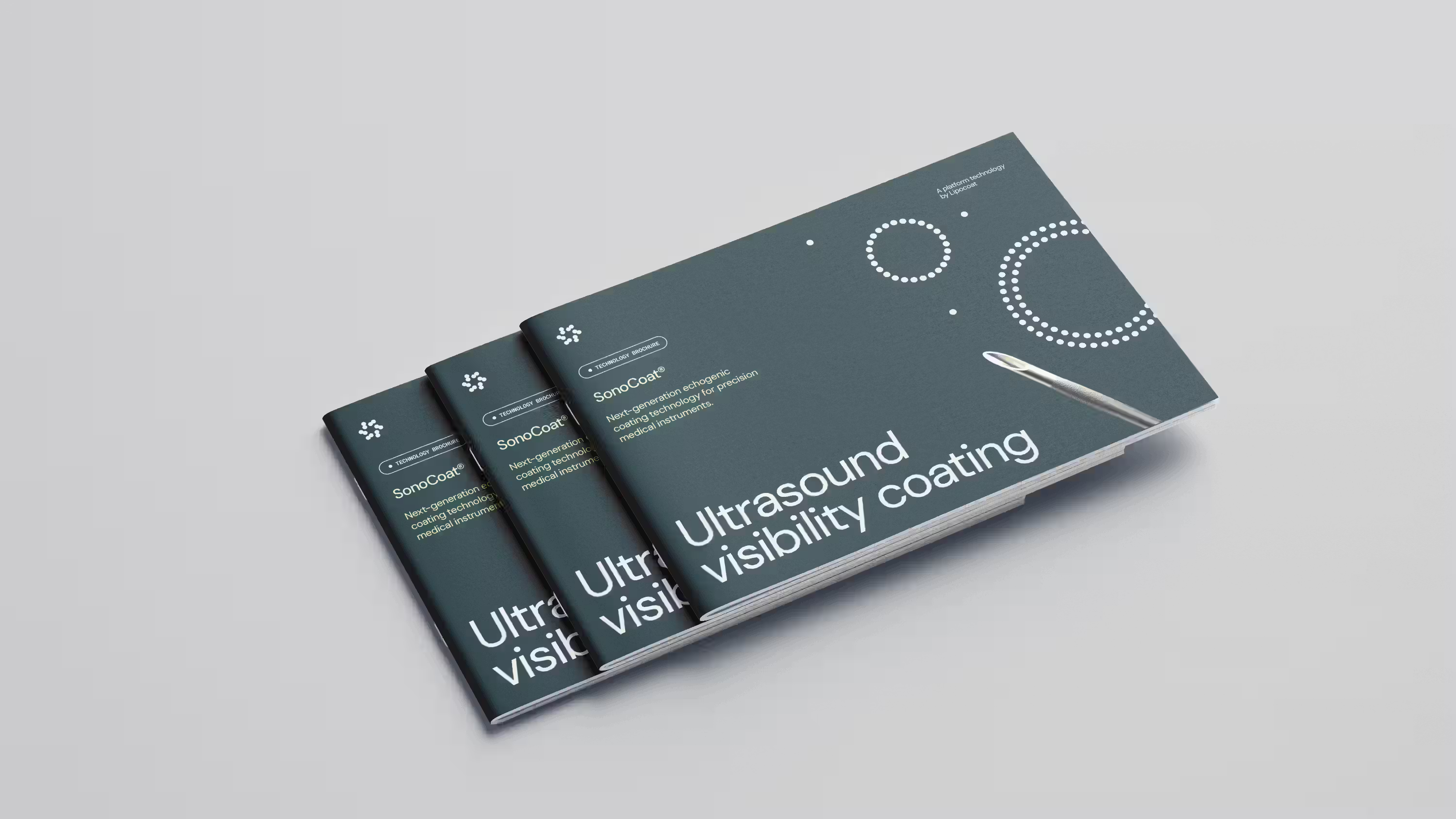
Real-time ultrasound guidance is now standard for CVC placement, yet needles still vanish at steep insertion angles. What SonoCoat® changes in vascular access, biopsy and regional anesthesia.