SonoCoat® Technology Featured in Scientific Publications
Independent peer-reviewed research
Bram Pape
September 16, 2026


Claims about ultrasound visibility are easy to make and hard to verify. The most useful evidence comes from research groups with no commercial interest in the outcome. SonoCoat® has been evaluated in four peer-reviewed publications by independent teams across three countries. Here is what each study set out to do, and where the coating appears in it.
Echogenic coatings are judged on one thing: whether a clinician can still see the instrument when the imaging conditions turn against them. Four independent research groups have put SonoCoat® through that test, in settings ranging from endoscopic ultrasound to prostate brachytherapy. The studies below were designed, run and published by those teams, not by us.
Tang, Vilmann et al., Gastrointestinal Endoscopy, 2016
The EUS Needle Identification Comparison and Evaluation study compared ten 22-gauge EUS-FNA needles: eight commercially available devices including SonoTip Pro Control, Expect Slimline, EchoTip Ultra, ClearView and EZ Shot 2, plus two prototype needles coated with SonoCoat®. Standardized, unedited videos were assessed by 43 EUS endoscopists and 17 radiologists, all blinded and unfamiliar with the devices. One SonoCoat®-coated prototype was rated best overall, ranking 10% to 40% higher than every other needle (P < .01), with no meaningful difference between how endoscopists and radiologists scored it. The authors also noted that the two needles coated with echogenic polymers were the only ones where the entire needle, rather than the tip alone, remained visible.
Brost, Stish, Lee, Urban and Deufel, Medical Physics, 2023
A team at the Mayo Clinic applied SonoCoat® to 17-gauge titanium high-dose-rate brachytherapy needles in three coating thicknesses (27, 40 and 64 µm) over a 2.5 cm region from the tip. The needles were imaged with a transrectal probe in a tissue-equivalent prostate phantom and in a cadaveric male pelvis, under conditions that deliberately reproduced the hardest cases: multiple stacked needles, acoustic shadowing and angled implants. The 64 µm coating reduced reverberation artifacts by 31 ± 14% and comet tail artifacts by 40% to 70%, and improved needle contrast at angles up to 30 degrees. Artifact reduction increased with coating thickness. The coated needles stayed high-contrast and well defined even when acoustically shadowed by other needles, which is the situation where digitizing needle positions normally fails.
Hopstaken, de Jong and Fütterer, CardioVascular and Interventional Radiology, 2021
Radboud University Medical Center tested three Sono-Sheath™ prototypes, each a polymeric sheath carrying a different version of the SonoCoat® coating at the tip, against a conventional liver biopsy needle with a sandblasted echogenic tip. Imaging was performed in a human cadaver at two insertion angles (30 and 60 degrees) and two depths (5 and 10 cm), with videos blinded and scored on Likert scales by five independent radiologists, alongside quantitative contrast measurement in a phantom. All three prototypes outranked the control needle at 60 degrees and 5 cm depth, and matched it elsewhere. The radiologists reported more confidence taking a biopsy with the echogenic sheaths. The phantom analysis showed a statistically significant effect of the sheath on echogenic intensity (p = 0.004).
Hovgesen, Wilhjelm, Vilmann and Kalaitzakis, Journal of Ultrasound in Medicine, 2022
This PRISMA systematic review from the Technical University of Denmark and Herlev University Hospital screened the published literature and identified 24 papers reporting visibility measures for 33 different echogenic needles. The authors grouped the needles by surface characteristic, coated versus dimpled versus standard, and ranked them. Echogenic needles outperformed standard needles, particularly at steep angles. Among the echogenic group, coated needles appeared to be better visualized in vivo than non-coated, dimpled designs, with SonoCoat® outranking the EchoTip® in the study it appeared in. The authors are careful here: no single paper compared all the leading needles head to head, so they draw no definitive winner. They do argue that smooth echogenic coatings are worth developing further, because added surface roughness raises concerns around needle tract seeding.
Different specialties, different imaging setups, different countries, and one recurring finding: the advantage of the coating grows as the imaging gets harder. Steep angles, greater depth, acoustic shadowing from neighboring instruments. Those are precisely the conditions under which a conventional needle stops being useful on screen.

LipoCoat and SonoCoat have earned recognition from leading institutions and regulatory bodies. Our coatings meet the highest standards for biocompatibility and clinical efficacy.
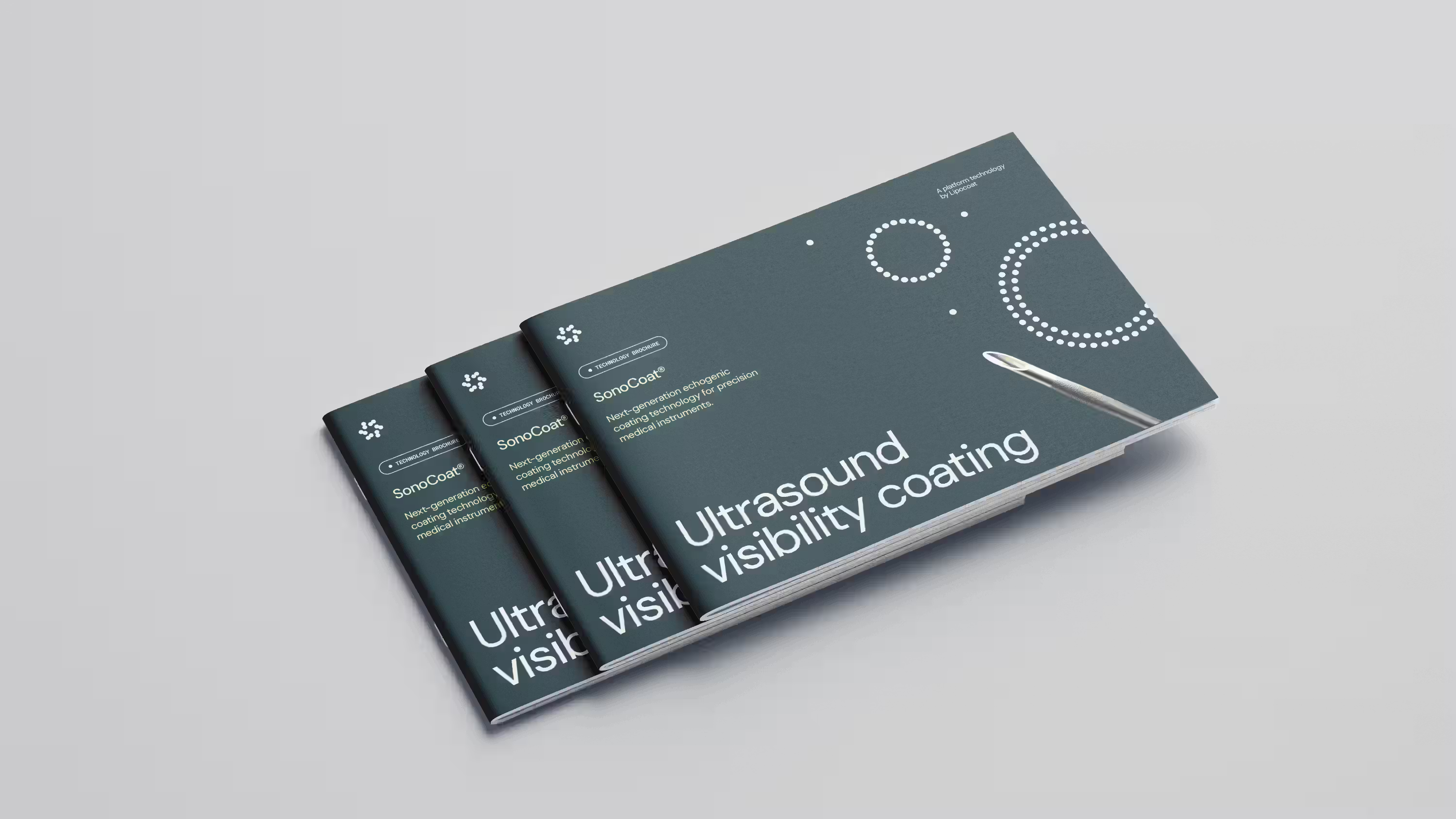
Real-time ultrasound guidance is now standard for CVC placement, yet needles still vanish at steep insertion angles. What SonoCoat® changes in vascular access, biopsy and regional anesthesia.