Publication

SonoCoat® Needles Tested by Combat Medics

Nine combat medics, one post-mortem model and an echogenic needle: results of a Journal of Vascular Access study.
Fabian Stein
October 23, 2026

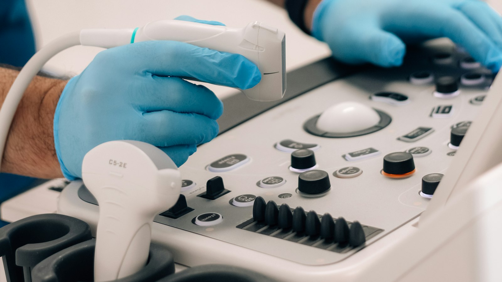
Gloved hands operating an ultrasound console with a probe

Introduction

A proof-of-concept study in the Journal of Vascular Access tested SonoCoat® echogenic needles with nine combat medics on a pressurized post-mortem model. Needle shaft visibility scored 4.0 against 2.0 for a conventional needle. Procedure time did not differ significantly.

Key takeaways

  • Nine Special Operation Forces combat medics with no ultrasound experience obtained femoral arterial access on a pressurized post-mortem model. All nine succeeded, within 3 minutes.
  • Needle shaft visibility received a median score of 4.0 with a SonoCoat® needle against 2.0 with a conventional needle (scale 1–5, p = 0.02).
  • Procedure time did not differ significantly between needle types (p = 0.57).
  • This is a proof of concept with nine participants. The authors calculate that a follow-up study needs about 28.

Why needle visibility matters

Femoral arterial access is the first step for REBOA (resuscitative endovascular balloon occlusion of the aorta), a temporary bridge to definitive bleeding control in non-compressible torso hemorrhage. In hemorrhagic shock, arteries and veins contract, which makes them hard to find. Medics in the field therefore need to obtain this access under difficult conditions.

Ultrasound guidance is widely used for vascular access. A meta-analysis of seven randomized trials with 546 patients (Gu et al., Critical Care 2014) found a first-attempt success rate of 48.5% with ultrasound-guided radial artery catheterization against 30.7% without (RR 1.55; 95% CI 1.02–2.35). The authors rated all trials at high risk of bias and the heterogeneity as high, so the result calls for careful reading.

Guidance works only when the needle can be followed on screen. A smooth needle surface reflects sound mainly at near-perpendicular incidence. Above roughly 45 degrees, very little signal returns. Deeper vessels require steeper angles, and the needle fades from the image.

SonoCoat® addresses this at the surface. The coating contains embedded microspheres that scatter the ultrasound beam in all directions. It is applied to the finished instrument, so the needle material itself stays unchanged.

The study in brief

  • Publication: Van de Voort et al., Journal of Vascular Access 2025;26(3):1016–1023, DOI 10.1177/11297298241256171
  • Institutions: Alrijne Hospital (Leiderdorp), Erasmus MC (Trauma Research Unit and Skillslab, Rotterdam), Defense Healthcare Organisation (Utrecht)
  • Design: Proof of concept, blinded to the order of needle types
  • Participants: 9 combat medics with no ultrasound or endovascular experience
  • Training: Four steps: theory, radial artery, REBOA Access Task Trainer, final test
  • Final test: Puncture of the common femoral artery on a pressurized post-mortem human specimen
  • Needle: 18G, 7 cm, SonoCoat® over 27.5 mm of the distal shaft. The conventional needle carried a non-echogenic dummy coating
  • Measures: Time to pulsatile water flow, self-ratings (1–5) and observer ratings (0–5)

The primary aim was to test a microteaching program. The echogenic needle was a secondary question.

What the medics found

  • Success. All nine participants obtained femoral arterial access in the specimen.
  • Visibility. The shaft scored 4.0 with the echogenic needle and 2.0 with the conventional needle (p = 0.02). Shaft identification also scored higher (4.0 versus 2.0, p = 0.02).
  • Needle tip. Differences in tip visibility and identification were not significant in the overall comparison (p = 0.16 and p = 0.09). In the second attempt, tip identification scored higher with the echogenic needle (4.0 versus 2.0, p = 0.048).
  • Preference. Seven of nine participants (78%) preferred the echogenic needle.
  • Time. Procedure time did not differ significantly between needle types (p = 0.57). As a group, participants were significantly faster in the second attempt (p = 0.004). In the second attempt the median time was 51 seconds with the echogenic needle and 55 seconds with the conventional needle.

How to read the results

The study gives a first signal on visibility. Patient outcomes remain untested, because no patients took part.

  • The groups were small: four or five participants per needle type per attempt.
  • In the first attempt the median time with the echogenic needle was higher (149 versus 113.5 seconds). The difference between needle types is not significant in the overall analysis.
  • The specimen deteriorated after about 25 puncture attempts.
  • The test took place under controlled conditions, without the light, noise and stress of the field.
  • First-pass success and complications were not reported as outcomes.

The next step is a larger study with simulated austere conditions. The authors expect that the preference for the coated needle may grow under poor light, noise and exhaustion. That remains a hypothesis.

What other research shows

The authors place their result next to three other studies:

  • Liver biopsy in post-mortem specimens. Radiologists were more confident with an echogenic sheath than with the control needle at a 30° insertion angle. The sheath had a significantly higher echogenic intensity, without artifacts or shadowing (Hopstaken et al.).
  • Phantom study. Echogenic needles showed higher objectively measured and subjectively experienced visibility than conventional needles (Nakagawa et al.).
  • Observational study with 69 inexperienced participants. Echogenic needles gave no objectively measured performance improvement over conventional needles (Phelan et al.).

The picture is mixed. Visibility improves in several models. An objective performance gain is not yet shown.

What this means for device manufacturers

A coating applied to an existing instrument after production adds visibility without changing the device design.

  • Materials: stainless steel, nitinol, titanium, ceramics and polymers such as PU, nylon, PEBAX, PEEK, PVC and polysilicones.
  • Layer thickness: 35–90 µm, tuned to the acoustic requirements of the application.
  • Applications: needles for regional anesthesia, biopsy and vascular access, catheters (CVC, PICC) and interventional radiology.
  • Safety: the coating has passed ISO 10993 testing and contains no drug or biocide, according to the SonoCoat® product page.
  • Earlier evidence: in the NICE study, 60 ultrasound specialists blind-rated videos of ten 22G EUS-FNA needles. The SonoCoat® needle scored highest overall (Gastrointestinal Endoscopy 2016;84(3):424–433). More studies are summarized in SonoCoat® Technology Featured in Scientific Publications.

Whether a change to an existing product requires a notification or a new submission depends on the product and the scope of the change. The manufacturer's regulatory team assesses that.

Want to evaluate SonoCoat® on your own needle or catheter? Request a feasibility study and get real performance data for your device.

Transparency

The authors declare no conflicts of interest. The first and corresponding author are affiliated with Alrijne Hospital. SonoCoat® is a Lipocoat Group technology.


References

  • Van de Voort JC, Stark PW, van Dongen TTCF, Borger van der Burg BLS, Hoencamp R. Ultrasound guided arterial access for combat medics: a blinded proof-of-concept study using echogenic needles. J Vasc Access 2025;26(3):1016–1023. DOI 10.1177/11297298241256171
  • Gu WJ, Tie HT, Liu JC, et al. Efficacy of ultrasound-guided radial artery catheterization: a systematic review and meta-analysis of randomized controlled trials. Crit Care 2014;18:R93. Springer
  • Lipocoat Group. SonoCoat® ultrasound visibility

Evaluate SonoCoat® on your device

Talk to our team about a feasibility study on your device.
Updates

Also interesting

Clinician in a blue surgical gown holding a medical endoscope next to a monitor during an endoscopic procedure
Grant Project

BILECOAT Develops a biliary stent coating to keep stents open

BILECOAT develops a LipoCoat® lipid coating for biliary stents with the University of Twente. The goal is a stent that stays open longer and needs fewer repeat procedures.
Black and white MRI scan images shown side by side
Grant Project

MIME Receives Health~Holland MKB Call Funding

MIME develops a SonoCoat® coating that makes medical devices visible in ultrasound, CT and MRI, together with Radboud University Medical Center. Funded by the Health~Holland MKB Call.
Colorful bacterial colonies growing on a dark culture surface
Grant Project

UROCOAT Receives Health~Holland MKB Call Funding

UROCOAT develops an antibiotic-free LipoCoat® coating for urinary catheters with the University of Twente. The target is to reduce catheter-associated infections by 50–90%. Funded by the Health~Holland MKB Call.